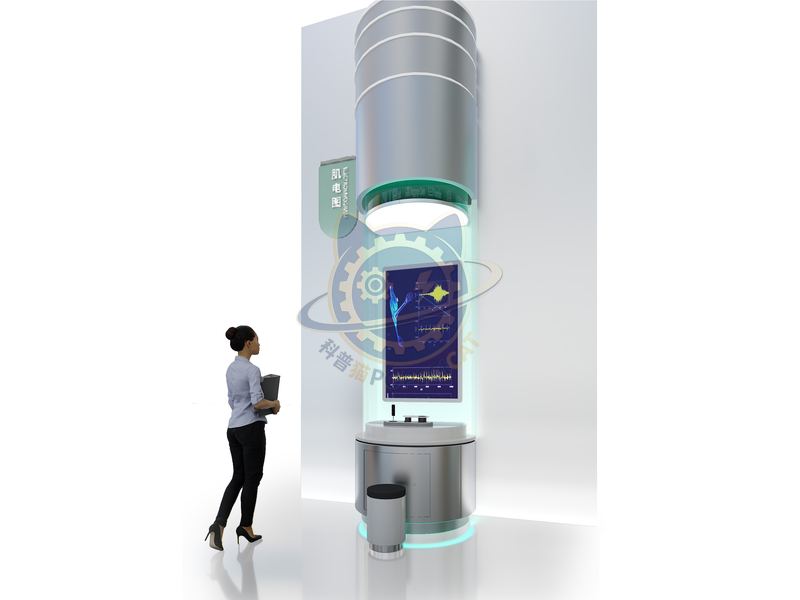
Electromyography is a muscle bioelectric pattern recorded by an electromyography device.
| Category | Life Science & Health |
|---|---|
| Standard size | Custom size |
| Supply | Single unit or batch · OEM to your drawing |
| Lead time | Standard 4–8 weeks after deposit (confirmed per contract) |
| Warranty | 1 year, structure and control |
| Documents | Specification sheet · control diagram · spare-part list |
A standard catalogue model: the dimensions, control package and finish are fixed, which is what keeps the price and the lead time predictable. Size, colour and interface language can be adapted without a new design cycle.
Standard models from this range are also listed in our online store, priced in USD — see the price yourself ↗
Electromyography is a muscle bioelectric pattern recorded by an electromyography device. The principle is to detect and record the weak electrical signals generated by muscles during contraction and relaxation. Electromyography is widely used to diagnose the health status of nerves and muscles, and is likened to a "miracle tool" that can make nerves and muscles "speak".
The unit is designed for unsupervised public use: controls are recessed or shielded, moving parts are enclosed, and the interface is operable by a child of about six with no instruction. Where a screen is used, the language can be switched without changing the hardware, and the content can be replaced on site.
Electromyography is a test that records the electrical activity of the nervous muscles and is the main means of examining the functional status of the peripheral nervous system. The main examination methods include nerve transmission examination and needle electrode electromyography examination, which record the electrical signals actively or induced by muscles and nerves. Skeletal muscles undergo potential changes during excitation due to the conduction and diffusion of action potentials in muscle fibers. The graph obtained by guiding and recording the potential changes that occur during skeletal muscle excitation using appropriate methods is called electromyography.
Every unit leaves the factory after a running test: mechanisms cycled, electronics burned in, edges dressed, fixings torqued and marked. Wear parts — bearings, sliders, lamps, belts, touch panels — are modular and listed on the spare-part sheet, so a venue technician can replace them without returning the exhibit. Crating is plywood with foam blocking; the loading plan and packing list travel with the shipment.
Other models from the Life Science & Health line, all held to the same specification and the same control package.

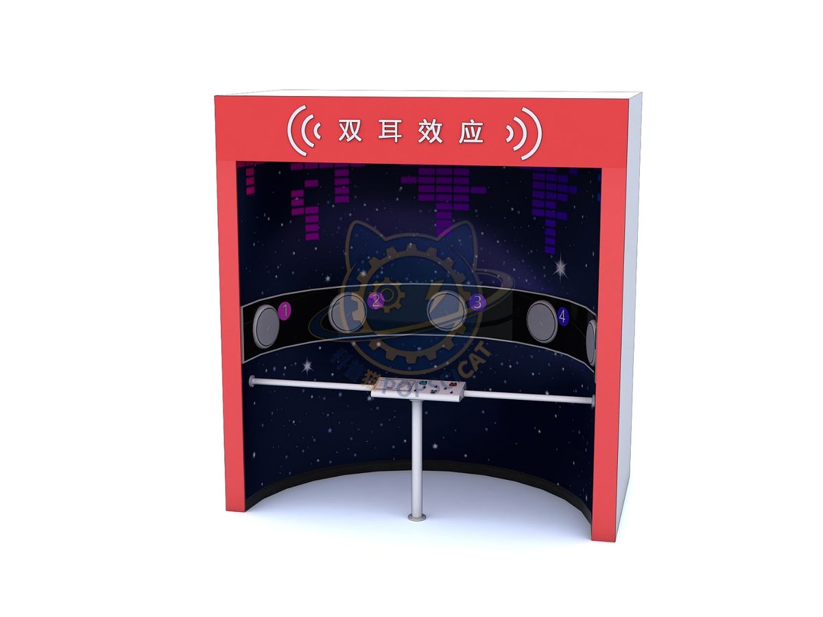
Sound reaches two ears at different times; visitors point to the source.

An adjustable lens projects a real image onto a retina screen.

Projected anatomy on a body-sized screen layer by layer.

Contract a model biceps and watch the tendon pull the forearm.
Attach a theme outline, a floor plan, or nothing at all — a photo of the space is enough to start. Quotations are issued against a real exhibit list, not per-square-metre guesses.
Scan to add us