Ultrasonic imaging

Ultrasound is a type of mechanical wave that is used in medicine to image human tissues through wave scattering (the…

Ultrasonic imaging — Life Science & Health exhibit for science museums
Reference image
CategoryLife Science & Health
Standard sizeCustom size
SupplySingle unit or batch · OEM to your drawing
Lead timeStandard 4–8 weeks after deposit (confirmed per contract)
Warranty1 year, structure and control
DocumentsSpecification sheet · control diagram · spare-part list

A standard catalogue model: the dimensions, control package and finish are fixed, which is what keeps the price and the lead time predictable. Size, colour and interface language can be adapted without a new design cycle.

Ask about this model → Email us

Standard models from this range are also listed in our online store, priced in USD — see the price yourself ↗
01 / Overview

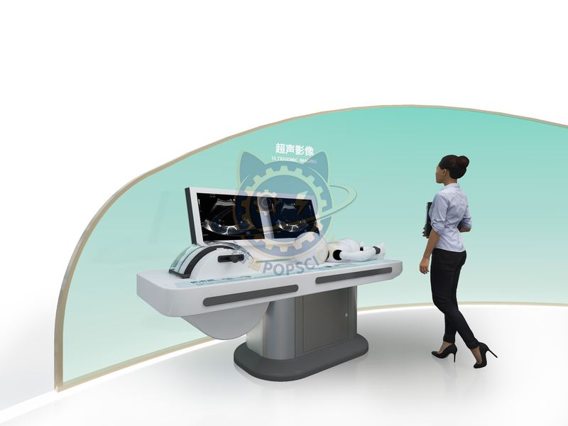
Ultrasound is a type of mechanical wave that is used in medicine to image human tissues through wave scattering (the phenomenon of sound waves scattering outwards when encountering uneven structures), known as ultrasound and color ultrasound. There are differences in the speed and density of sound in different tissues and organs of the human body. After the sound waves are emitted by the ultrasonic probe and transmitted to the human body, they will produce strong and weak reflection and scattering waves at the interface and uneven areas inside the organs, and finally return to the transducer on the surface of the body (the core component responsible for emitting and receiving sound waves inside the probe). Ultrasound examination requires the application of a coupling agent, which can eliminate the air between the probe and the skin, thereby improving the clarity of image display. Ultrasound diagnosis includes vascular ultrasound, cardiac ultrasound, abdominal ultrasound (visceral), thyroid ultrasound, breast ultrasound, obstetric and gynecological ultrasound, etc.

02 / How visitors use it

The unit is designed for unsupervised public use: controls are recessed or shielded, moving parts are enclosed, and the interface is operable by a child of about six with no instruction. Where a screen is used, the language can be switched without changing the hardware, and the content can be replaced on site.

03 / The science behind it

The physical characteristics of ultrasound: the higher the frequency, the higher the resolution, but the weaker the penetration power; The lower the frequency, the better the penetration, but the poorer the resolution. In clinical applications, ultrasound requires high resolution (high frequency) and sufficient penetration depth to see deeper structures (low frequency) in order to obtain clear images. Therefore, it is necessary to choose the optimal frequency. If the frequency is too high, the penetration depth of ultrasound may not be sufficient, and the desired structure may not be achieved; If the frequency is too low, ultrasound will penetrate deeper than necessary, and unnecessary low frequencies will produce blurry low resolution images.

04 / Built for a public floor

Every unit leaves the factory after a running test: mechanisms cycled, electronics burned in, edges dressed, fixings torqued and marked. Wear parts — bearings, sliders, lamps, belts, touch panels — are modular and listed on the spare-part sheet, so a venue technician can replace them without returning the exhibit. Crating is plywood with foam blocking; the loading plan and packing list travel with the shipment.

Same category

More in Life Science & Health

Other models from the Life Science & Health line, all held to the same specification and the same control package.

Start a project

Send us your exhibit list.
We will come back with a price.

Attach a theme outline, a floor plan, or nothing at all — a photo of the space is enough to start. Quotations are issued against a real exhibit list, not per-square-metre guesses.

WeChat QR code — scan to message us Scan to add us
on WeChat